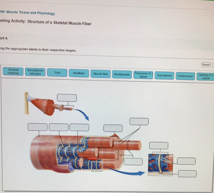
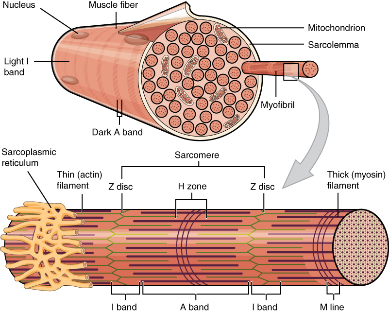
43 art-labeling activity: structure of a skeletal muscle fiber
chapter 9 Anatomy Flashcards | Quizlet Art-labeling Activity: The structure of a skeletal muscle fiber PICTURE Chapter Test - Chapter 9 Question 3 Which thin-filament-associated structure is distinguished by its constituents of three globular subunits, one of which has a receptor that binds two calcium ions? a) G-actin b) nebulin c) tropomyosin d) troponin D ... AP 1- CHAPTER 9 MASTERING ASSIGNMENTS Flashcards | Quizlet Art-labeling Activity: The structure of a skeletal muscle fiber PICTURE Which thin filament-associated protein binds two calcium ions? troponin Action potential propagation in a skeletal muscle fiber ceases when acetylcholine is removed from the synaptic cleft.
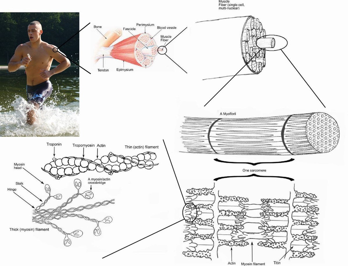
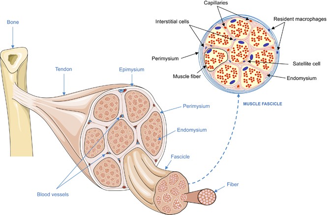
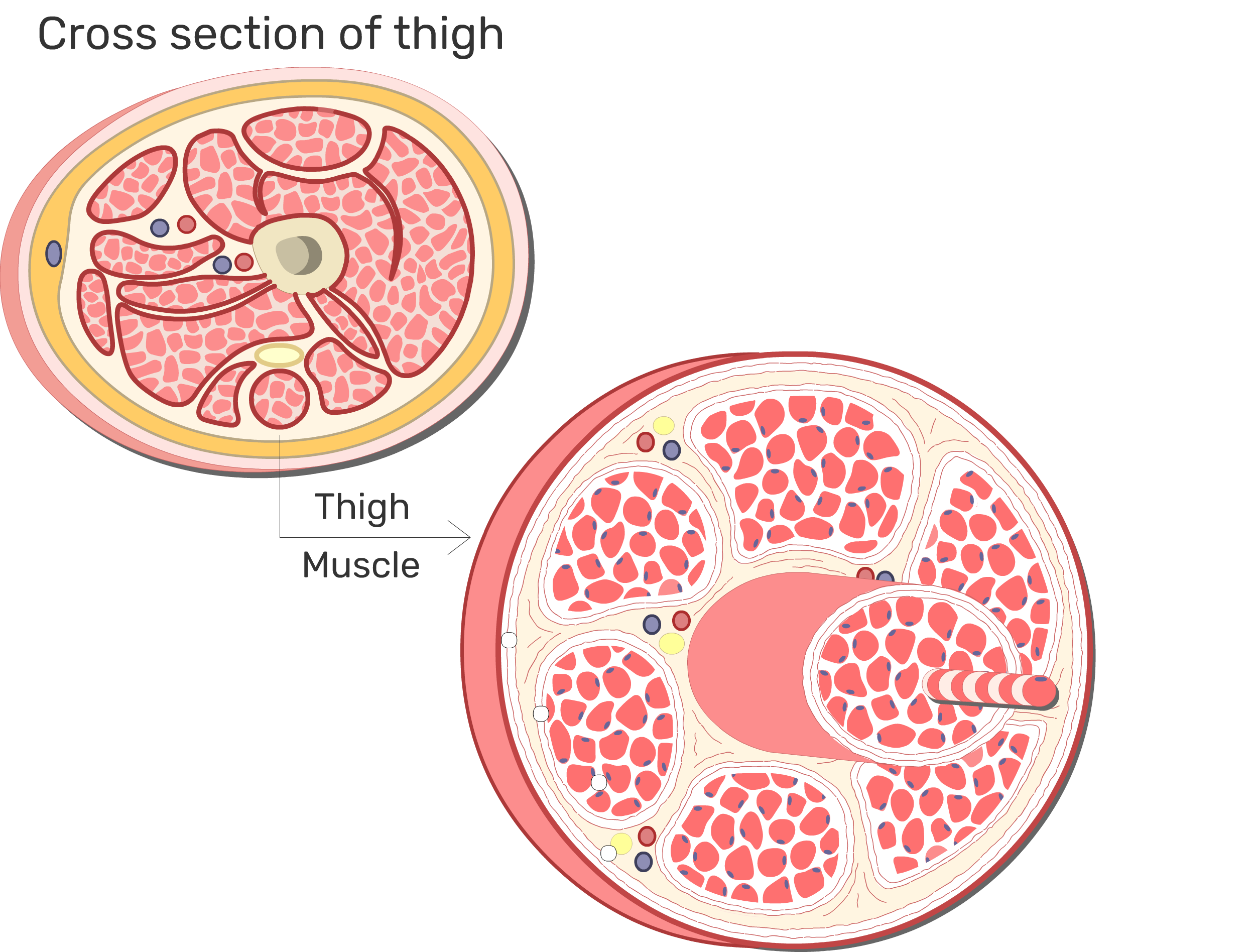
Art-labeling activity structure of a skeletal muscle fiber The structure of a skeletal muscle fiber is attached. What is the composition of a muscle fiber? Muscle is contractile tissue that is organized into coordinated systems for maximum efficiency. Muscle systems in humans are classified based on their gross appearance and cell location.

Art-labeling activity: structure of a skeletal muscle fiber
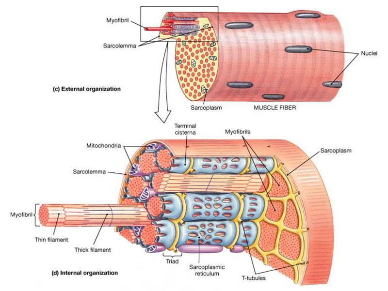
11.2: Microscopic Anatomy of Skeletal Muscles - Home - Biology LibreTexts Microscopic Anatomy of Skeletal Muscles. Skeletal muscle is found attached to bones. It consists of long multinucleate fibers. The fibers run the entire length of the muscle they come from and so are usually too long to have their ends visible when viewed under the microscope. The fibers are relatively wide and very long, but unbranched. 10.2 Skeletal Muscle - Anatomy and Physiology 2e | OpenStax 10.1Overview of Muscle Tissues 10.2Skeletal Muscle 10.3Muscle Fiber Contraction and Relaxation 10.4Nervous System Control of Muscle Tension 10.5Types of Muscle Fibers 10.6Exercise and Muscle Performance 10.7Cardiac Muscle Tissue 10.8Smooth Muscle 10.9Development and Regeneration of Muscle Tissue Key Terms Chapter Review Skeletal Muscle Fiber Structure and Function - Open Textbooks for Hong Kong A muscle fiber is composed of many fibrils packaged into orderly units. The orderly arrangement of the proteins in each unit, shown as red and blue lines, gives the cell its striated appearance. The striated appearance of skeletal muscle tissue is a result of repeating bands of the proteins actin and myosin that occur along the length of ...
Art-labeling activity: structure of a skeletal muscle fiber. Art-labeling Activity: The Structure of a Skeletal Muscle Fiber Art-labeling Activity: The Structure of a Skeletal Muscle Fiber + − Learn Test Match Created by BabeRuthless0504 Terms in this set (2) Art-labeling Activity: The Structure of a Skeletal Muscle Fiber ... Art-labeling Activity: The Structure of a Skeletal Muscle Fiber ... Students also viewed Ch 10 lab map 23 terms mnalley09 Plus chapter 9 Flashcards | Quizlet Art-labeling Activity: The structure of a skeletal muscle fiber PICTURE Chapter Test - Chapter 9 Question 3 Which thin-filament-associated structure is distinguished by its constituents of three globular subunits, one of which has a receptor that binds two calcium ions? a) G-actin b) nebulin c) tropomyosin d) troponin D ... Question: Art-labeling activity: structure of skeletal muscle fiber ... Art-labeling activity: structure of skeletal muscle fiber.Drag the appropriate lablels to their respective targets. This problem has been solved! You'll get a detailed solution from a subject matter expert that helps you learn core concepts. See Answer Solved Art-labeling Activity: The Structure of a Sarcomere - Chegg Question: Art-labeling Activity: The Structure of a Sarcomere Part A Drag the labels to the appropriate location in the figure. Reset Help A band Barmere Hand band MI Art-labeling Activity: The structure of a skeletal muscle fiber Part A Drag the labels onto the diagram to identity structural features associated with a skeletal muscle fiber.
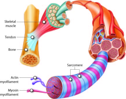
chapter 9 Flashcards | Quizlet Art-labeling Activity: The structure of a skeletal muscle fiber PICTURE Chapter Test - Chapter 9 Question 3 Which thin-filament-associated structure is distinguished by its constituents of three globular subunits, one of which has a receptor that binds two calcium ions? a) G-actin b) nebulin c) tropomyosin d) troponin D ... 10.2 Skeletal Muscle The striations of skeletal muscle are created by the organization of actin and myosin filaments resulting in the banding pattern of myofibrils. These actin and myosin filaments slide over each other to cause shortening of sarcomeres and the cells to produce force. Interactive Link Questions 10.5 Types of Muscle Fibers - Anatomy and Physiology | OpenStax Want to cite, share, or modify this book? This book uses the Creative Commons Attribution License and you must attribute OpenStax. Attribution information. If you are redistributing all or part of this book in a print format, then you must include on every physical page the following attribution: Muscle Fibers: Anatomy, Function, and More - Healthline Muscle fibers can be found in skeletal, cardiac, and smooth muscles, and work to do different things in the body. ... ST fibers are good for long lasting activities. These can include things like ...
structure of skeletal muscle fiber Flashcards | Quizlet actin and myosin Epimysium covers the entire skeletal muscle Perimysium The connective tissue that surrounds fascicles. Endomysium Surrounds individual muscle fibers Sarcolemma plasma membrane of a muscle fiber Sarcoplasm cytoplasm of a muscle cell sarcoplasmic reticulum specialized endoplasmic reticulum of muscle cells terminal cisternae BIO 200 Chapter 9 - Muscle Tissue Physiology Flashcards | Quizlet Art-Labeling Activity: Structure of a Skeletal Muscle Fiber When the sarcomere contracts and shortens__________. the A band stays the same The storage and release of calcium ions is the key function of the: sarcoplasmic reticulum. A group of skeletal muscle fibers together with the surrounding perimysium form a (n): fascicle. Skeletal Muscle Fiber Structure and Function - Open Textbooks for Hong Kong A muscle fiber is composed of many fibrils packaged into orderly units. The orderly arrangement of the proteins in each unit, shown as red and blue lines, gives the cell its striated appearance. The striated appearance of skeletal muscle tissue is a result of repeating bands of the proteins actin and myosin that occur along the length of ... 10.2 Skeletal Muscle - Anatomy and Physiology 2e | OpenStax 10.1Overview of Muscle Tissues 10.2Skeletal Muscle 10.3Muscle Fiber Contraction and Relaxation 10.4Nervous System Control of Muscle Tension 10.5Types of Muscle Fibers 10.6Exercise and Muscle Performance 10.7Cardiac Muscle Tissue 10.8Smooth Muscle 10.9Development and Regeneration of Muscle Tissue Key Terms Chapter Review
BIO 200 Chapter 9 - Muscle Tissue Physiology Flashcards ...
11.2: Microscopic Anatomy of Skeletal Muscles - Home - Biology LibreTexts Microscopic Anatomy of Skeletal Muscles. Skeletal muscle is found attached to bones. It consists of long multinucleate fibers. The fibers run the entire length of the muscle they come from and so are usually too long to have their ends visible when viewed under the microscope. The fibers are relatively wide and very long, but unbranched.
Solved Art-labeling activity: structure of skeletal muscle ...
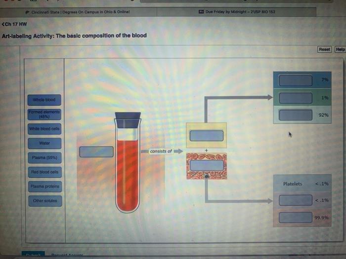
Answered: 1% Whole tiood Farmed elemente (45%)… | bartleby

A) Illustration of skeletal muscle structure copied with ...

The structure and function of cardiac t-tubules in health and ...
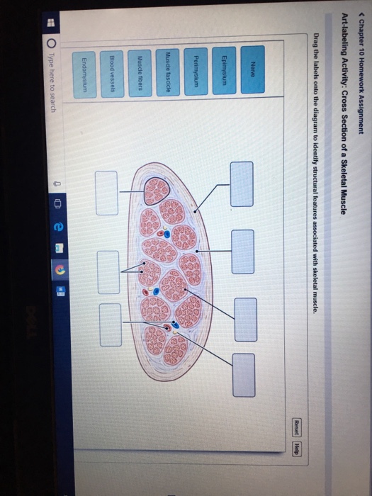
Solved Art-labeling Activity: Cross Section of a Skeletal ...
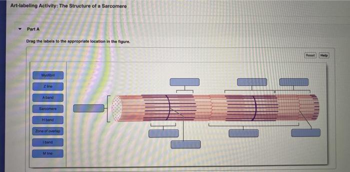
Solved Art-labeling Activity: The Structure of a Sarcomere ...

10 muscles
Muscle contraction - Wikipedia

Skeletal Muscle | Anatomy and Physiology I

Skeletal Muscle 4- Thin Filaments - YouTube

Pin on Chapter 11: Muscular Tissue

10.21aaa.png - Art-labeling Activity: Figure 10.21a Drag the ...

Quiz 10 Chapter 12 - CNS.pdf - 2/10/22, 10:58 PM Quiz 10 ...

T Lymphocyte-Captured DNA Network for Localized Immunotherapy ...

Muscular Levels of Organization | Anatomy and Physiology I ...

High percentage of fibers with central nuclei in fast-twitch ...
Cell death, clearance and immunity in the skeletal muscle ...
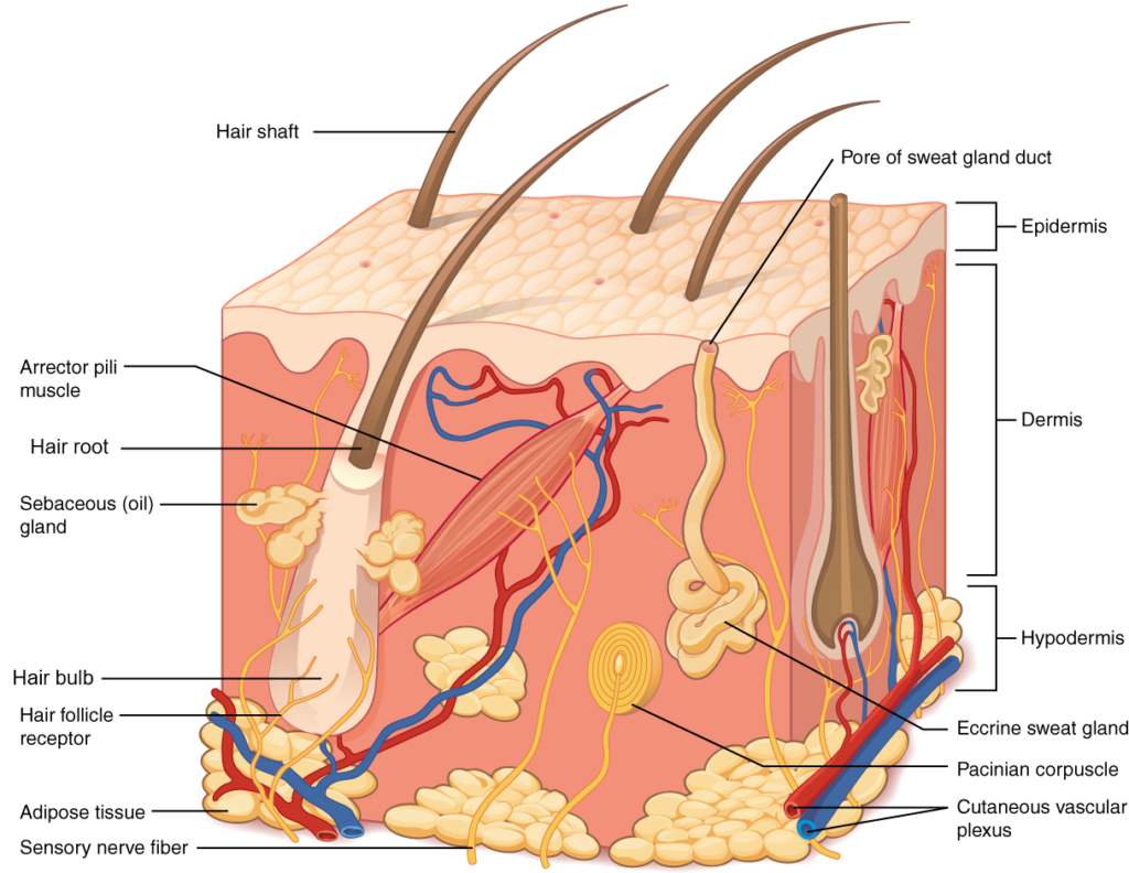
Unit 11: The Integumentary System – Douglas College Human ...

A&P 1- CHAPTER 9 MASTERING ASSIGNMENTS Flashcards | Quizlet
Frontiers | Metabolomic Profile of Skeletal Muscle and Its ...
Types of muscle cells: Characteristics, location, roles | Kenhub
Essentials of Anatomy and Physiology

Induced pluripotent stem cells for periodontal regeneration ...
Skeletal muscle fibers: arrangement and diagram | GetBodySmart

Ex 12 Microscopic Anatomy & Organization of Skeletal Muscle ...

Art Labeling Activity Figure 4.3 2 of 2.png - | Course Hero
Skeletal Muscle | Anatomy and Physiology | | Course Hero

10 Muscle Tissue. - ppt download

Advanced Biology – 12/2/14 Warm Up Muscle Contraction - ppt ...

Microscopic Anatomy of Skeletal Muscle Flashcards | Quizlet

BIO 200 Chapter 9 - Muscle Tissue Physiology Flashcards ...
Answered: Part A 4. Drag the labels to the… | bartleby

Ch 10 lab map Flashcards | Quizlet

Lab Exercise 12.pdf - 7/6/2021 Lab Exercise 12 Lab Exercise ...
Innervation: the missing link for biofabricated tissues and ...
Anatomy Exam 2 Flashcards - Easy Notecards

Label the following in a diagram of a skeletal muscle fiber ...

Brain structure and function related to headache: Brainstem ...
Untitled

Neurolemmocyte On Skeletal Muscle Model - Human Anatomy ...
Muscular Levels of Organization | Anatomy and Physiology I ...
Figure 19.1 Srructures found in skeletal muscle fibers (cells ...
Post a Comment for "43 art-labeling activity: structure of a skeletal muscle fiber"